
The DDI team looks forward to taking care of your imaging needs when you visit our center. The following is some information to help you prepare.
Download the DDI Patient Sign in Form, or fill it out at the time of your appointment.
Before your Visit
Your doctor will provide you with a completed, signed, DDI referral slip. It is very important that you bring this referral slip with you to your DDI appointment, as well as having it available when you call us for your appointment. It tells us exactly what types of images your doctor needs.
*Note: DDI cannot image a patient without a referral from a doctor.
Find the nearest DDI Imaging Center. Our locations are available on our Locations Page, as well as on your referral slip.
Contact us to schedule your appointment
While walk-ins can be accommodated, there may be a wait. It is always best to call us for an appointment. We will be asking for the information from the referral slip provided to you by your doctors office. You can find our phone numbers on the Contact Us Page.
Rescheduling
Life happens and we can help. If you need to reschedule your appointment, call us and we will get your appointment scheduled on a day more suitable for you.
Cancellation
Please call us if you need to cancel your appointment so that we can make the appointment available to others.
Checking in
When you arrive at our office please check in with the receptionist. They will collect your referral slip, ask you to fill out a short form and process your payment. Before we begin your imaging you will be asked to remove all forms of metal from your head.
Payment method
Payment is due at the time of your appointment. You will be provided a fee estimate when scheduling your appointment. We accept cash, checks and major debit/credit cards. If you have insurance, we will assist you with your claim form. Your carrier will reimburse you directly if they determine that the procedure is a covered benefit under your plan.
Arriving late
If you are going to be more that just a few minutes late, please call our office to let us know. We can usually manage, or we can reschedule you accordingly. If you arrive more than a few minutes late you may have to wait.
Your imaging
A x-ray technologist will call you back for your imaging session requested by your doctor. Once your imaging session is complete, we’ll ask you to wait just a few minutes while the computers complete the processing of the data.
*Note: Some of the X-ray procedures require the patient to be able to hold their head completely still for up to 30 seconds in order to get the best possible image quality. If you have a physical condition that may interfere with this, please let our staff know when scheduling the appointment.
Q & A: What is this image used for?
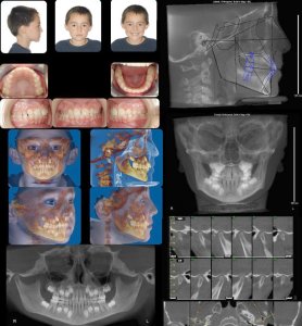
Oral Facial Photography can assist in explaining the face and teeth from different angles and are used to assist in diagnosis for bite, facial asymmetry and treatment options. Orthodontists often use these photographs to document treatment time- points like Before and After.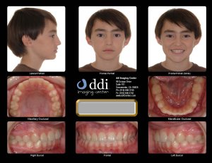
Full Mouth X-Rays are primarily used to identify dental cavities, short roots, and dental abnormalities involving the bone adjacent to the teeth.

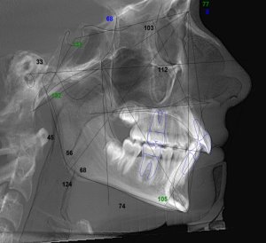
Cephalometric projection images Lateral and Posterior/Anterior view are used to demonstrate the bones and soft tissues of the head, and are helpful in evaluating facial growth. Measurements generated from digitizing the structures are used to determine the relationships between the upper and lower jaws, dental, skeletal, and soft tissues.

Panoramic image is an overview of the upper and lower jaws, erupted and un-erupted teeth, and it is used to evaluate the maxilla and mandible and can be used for root alignment, TMJ’s and sinuses.

Cone Beam CT is a 3 Dimensional digital scanner specifically designed to capture the face, teeth, airway, and jaws. Patients stay comfortably seated while the cone shaped beam rotates around 360 degrees within a matter of seconds. The volumetric data collected is formatted to produce 2D image projections as well as 3D volumes for treatment planning and diagnostic support.


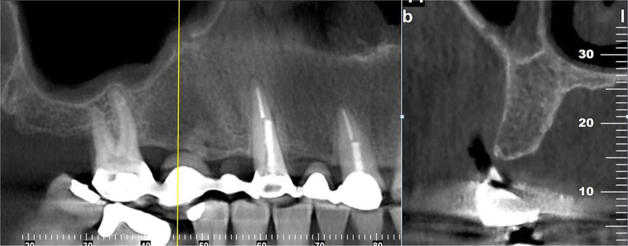
CBCT Implant scan uses special viewing software for visualizing if adequate bone is available prior to implant placement. The nerves in the lower jaw and the sinus in the upper jaw can be identified along with other relevant anatomy in the 3D scan for treatment planning.

CBCT Orthodontic scanning can provide the information required to perform a thorough pre-treatment analysis blueprint for braces, and clear aligner through the evaluation of the face, teeth, jaws, TMJ’s, and airway.
CBCT of the TMJ’s allows for the evaluation of Temporomandibular Joint disorder, and also diagnostic image of the teeth, sinus, jaws, and airway. Signs and symptoms of TMD may include pain in joint area as well as the face, ringing in the ears also know as tinnitus, and arthritis that may originate in many areas of the face and adjacent structures.

